For centuries, humanity has hungered to unravel the secrets of the microworld. From the first lenses crafted by Antonie van Leeuwenhoek to the intricate optical systems of the 19th century, scientists have desperately tried to peer into the hidden reaches of nature. Yet, by the early 20th century, physics hit a brick wall: the diffraction limit of light. Optical instruments were physically incapable of resolving objects smaller than the wavelength of visible light. The realm of viruses, the internal architecture of cells, and the very arrangement of atoms remained unreachable “black boxes,” reports toronto-future.com.
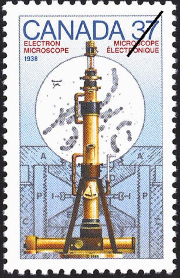
Everything changed in 1938. Within the walls of the McLennan Laboratory at the University of Toronto, two young graduate students, guided by an ambitious professor, finished work on a device that allowed humanity to gaze beyond the seemingly impossible. It was the first practical electron microscope in North America, and arguably the first truly operational one in the world. This breakthrough didn’t just launch the era of nanotechnology and modern virology; it remains a symbol of scientific triumph achieved despite the crippling financial constraints of the Great Depression.
The Birth of an Idea
By the start of the 20th century, physicists realized that because the wavelength of visible light is measured in hundreds of nanometers, any object smaller than half that length – like a virus or delicate cellular structure – would remain an invisible, blurry smudge. The solution came from quantum physics. Louis de Broglie hypothesized that electrons exhibit wave-like properties, and Hans Busch proved that magnetic fields could focus electron beams just as glass lenses focus light. Since an electron’s wavelength is thousands of times smaller than that of light, such a device could theoretically achieve mind-bending magnification.
The first prototype of an electron microscope was built by Ernst Ruska in Berlin in 1931. However, his apparatus was more of a physical demonstration than a functional tool: it frequently burned through samples, suffered from poor resolution, and was incredibly difficult to operate. This is exactly where the University of Toronto stepped onto the stage.
Professor Burton and His “Dream Team”
The visionary leader of the Canadian project was E.F. Burton, head of the physics department at the University of Toronto. Burton possessed uncanny scientific intuition. Having worked at the Cavendish Laboratory under J.J. Thomson (the discoverer of the electron), he deeply understood the potential of electron optics. In 1935, after attending a conference in Germany, Burton returned to Toronto with a firm intention: to build a microscope that would actually work for biologists and materials scientists.
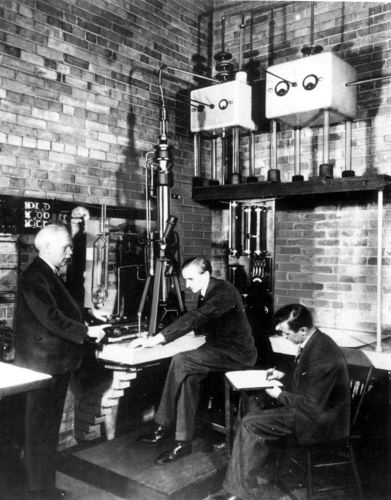
To turn this goal into reality, he recruited two talented students: Albert Prebus and James Hillier. Hillier was a master builder with the keen mind of an engineer, while Prebus brought a deep command of theoretical physics and mathematics. Together, they formed the perfect duo.
Building on a Shoestring: Vaseline and Rubber Bands
Work began immediately after Christmas in 1937. These were the lean years of the Great Depression, so funding was non-existent. Burton managed to secure only a few hundred dollars from the National Research Council. For perspective, modern developments of this caliber require millions in investments.
The graduate students had to be incredibly resourceful. Many parts were crafted by hand in the university workshops. Since the system had to operate in a high vacuum, sealing the joints proved difficult. Hillier and Prebus boiled their own vacuum grease, mixing raw rubber (sometimes just plain office rubber bands) with boiling Vaseline.
The technical hurdles were monumental:
- Power Stability. They needed to maintain 45,000 volts with a fluctuation of no more than one volt. Any jitter would blur the image completely.
- Cooling. A powerful electron beam generated so much heat that samples would simply evaporate.
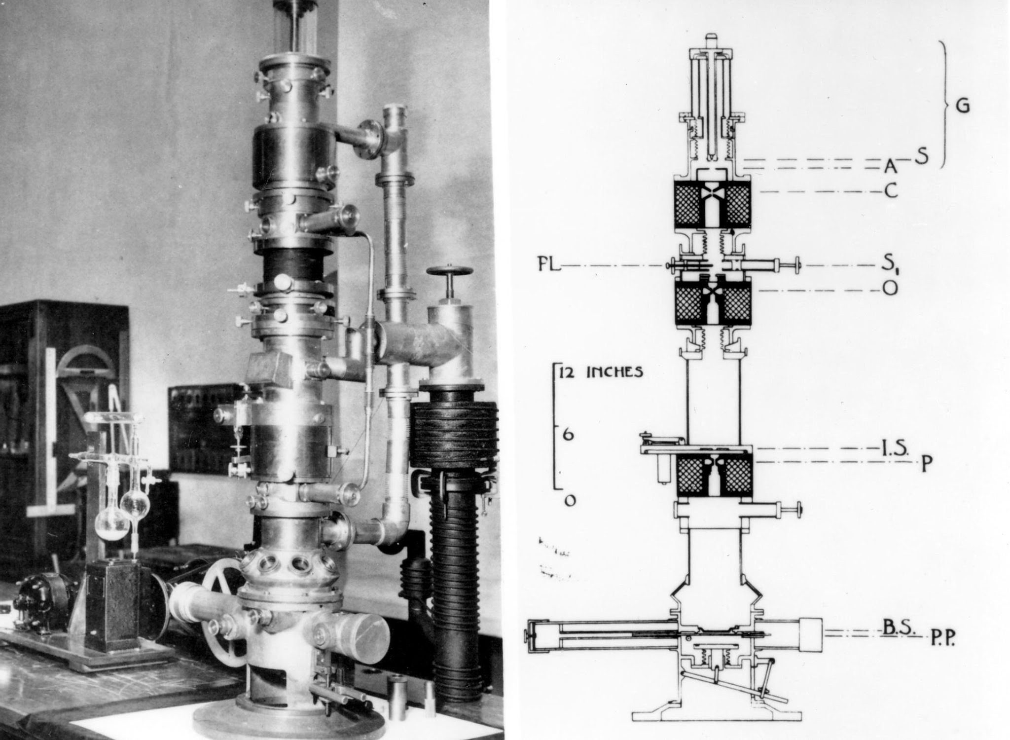
- Vacuum. The system consisted of six sections, and each had to be perfectly aligned and airtight.
Working double shifts – in the workshops during the day and on blueprints or assembly at night – the young scientists achieved the impossible. In just four months, by April 1938, they captured their first sharp images.
The 1938 Triumph
The 1938 model was a vertical column roughly two meters tall, mounted on a concrete slab to dampen vibrations. Unlike its German counterparts, the apparatus from Toronto produced a stable magnification of 10,000 to 30,000 times with a resolution far exceeding the limits of the best optical instruments of the era.
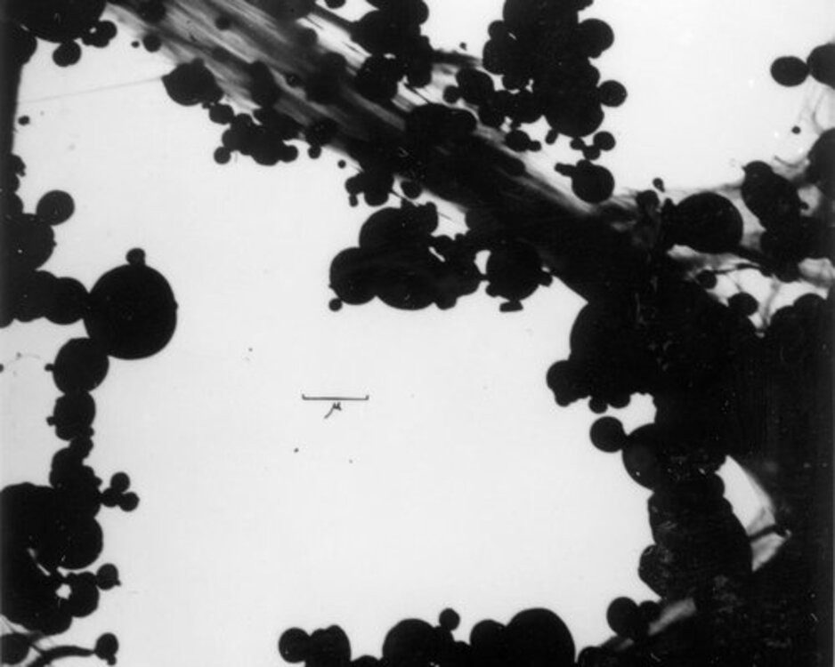
When the scientists demonstrated their images of bacteria and colloidal particles at a meeting of the American Physical Society in Toronto, the audience was stunned. It was akin to landing on another planet: a world that had previously existed only as theoretical models was suddenly visible.
One of Hillier’s colleagues, William Watson, recalled how difficult it was to convince the skeptics. Many American scientists believed electron microscopy was physically impossible and dismissed the first images as “fakes.” But the Toronto microscope worked flawlessly, proving its practicality day after day.
Science as Art: The “Nose Method”
Working with the first microscope demanded not just knowledge, but true craftsmanship, which Watson described as “art, not just science.” For example, preparing samples was an incredibly delicate affair. To create a thin support film (formvar), it had to be lifted off a glass plate. Often, the film would stick too tightly.
Watson devised a curious but effective method: he would run the glass plate along the side of his own nose to leave a microscopically thin layer of natural skin oil on the glass. After that, the film would lift off easily on the surface of the water. This technique was jokingly called “Watson’s Striptease,” and it remained a laboratory secret for many years.
War and the Manhattan Project
With the outbreak of the Second World War, the Toronto microscope became a strategic asset. Research was classified. The scientists worked on improving gas masks, studying how filter fibers trapped tiny aerosol particles.
However, there was a darker, more secretive chapter. In 1942, the laboratory was visited by renowned physicists George Paget Thomson and Francis Simon. Burton’s team was tasked with examining microscopic holes in metal meshes. Only after the war did it become clear these studies were part of the Manhattan Project – they concerned the gaseous diffusion separation of uranium isotopes. Thus, the invention from Toronto made its own contribution to ending the war.
Legacy and the Path Forward
Success in Toronto acted as a springboard for the project’s creators. James Hillier soon moved to the U.S., where he led the development of the first commercial electron microscopes at RCA. Thanks to his work, the technology born in a university lab became available to hospitals and research institutes worldwide.
Albert Prebus became a professor in Ohio, where he built an improved model of the microscope that served as the first operational unit in the United States.
The 1938 device stood in the hallway of the McLennan building for years as a monument to scientific courage. In 2010, it was transferred to the Canada Science and Technology Museum in Ottawa. Although it looks bulky and archaic compared to modern cryo-electron microscopes, it was this machine that paved the way for understanding DNA structure, investigating viruses (including COVID-19 decades later), and advancing nanotechnology.
Conclusion
The story of the electron microscope’s creation in Toronto is a testament to how a great idea can overcome a lack of resources. E.F. Burton, James Hillier, and Albert Prebus didn’t just assemble a complex instrument – they gifted humanity “new eyes.”
Today, when we observe individual atoms or protein structures, we owe it to those four months of intense labor in 1938. Back then, in that Toronto lab, the boundary of the invisible was crossed with the help of copper wire, Vaseline, and brilliant intuition. This microscope wasn’t just a Canadian achievement; it was the foundation upon which all modern science of the microworld is built.